Guo-Dong Huang1,
Jing Xiong2 ![]()
For correspondence:- Jing Xiong Email: xiongjing328@hotmail.com Tel:+8679188692962
Received: 11 May 2016 Accepted: 30 December 2016 Published: 25 February 2017
Citation:
Huang G, Xiong J.
A study on the ex
© 2017 The authors.
This is an Open Access article that uses a funding model which does not charge readers or their institutions for access and distributed under the terms of the Creative Commons Attribution License (http://creativecommons.org/licenses/by/4.0) and the Budapest Open Access Initiative (http://www.budapestopenaccessinitiative.org/read), which permit unrestricted use, distribution, and reproduction in any medium, provided the original work is properly credited..
Purpose: To investigate the ex
Methods: The ex
Results: MMP-9 and TIMP-1 were strongly expressed in the glandular epithelium and inflammatory cells. In addition, the ex
Conclusion: MMP-2, MMP-7, and MMP-9 are potential targets for therapeutic control of UC.
Introduction
Ulcerative colitis (UC) is a chronic inflammatory bowel disease that usually causes inflammation in the large intestine [1]. As an autoimmune disease, UC is influenced by environmental and genetic factors [2] and causes ulcers and inflammation in the colon [3]. The incidence of UC has increased gradually in recent years [4]; symptoms include bloody diarrhoea, fever, abdominal pain, and weight loss [5]. The pathogenesis of the disease is not fully understood, but a genetic risk is presumed [6]. The treatment of UC includes anti-inflammatory drugs and therapy targeted to the immune response [7,8].
Matrix metalloproteinases (MMPs) are proteolytic calcium-dependent enzymes that contain zinc and belong to the metzincin superfamily [9,10]. These enzymes play an important role in the degradation of extracellular matrix protein by cleaving its components [11]. MMPs are also involved in cell proliferation, adhesion, differentiation, and dispersion, and programed cell death [12]. Excreted by endothelial cells, neutrophils, macrophages, neutrophils, and lymphocytes [13,14], MMPs are controlled by tissue inhibitors of metalloproteinases (TIMPs) [15]. The balance between MMPs and TIMPs must be controlled to maintain and remodel cells and tissues. Imbalances in these proteins can lead to diseases such as tissue fibrosis, cardiovascular diseases, ulcers, and inflammation [15].
In this study, we evaluated the immunohistochemical associations of MMPs and TIMPS, specifically MMP-2, MMP-7, and MMP-9 and TIMP-1 and TIMP-2, respectively, in 50 patients with UC and their correlations with histochemical and pathological parameters. Increased understanding of the expression of these proteins in UC could provide novel therapeutic targets.
Methods
The study complied with National Institutes of Health guidelines [16] and was approved by Bioethics Committee of The First Affiliated Hospital of Nanchang University, Nanchang, Jiangxi, China (ref no. NU/FAH/BioEt/2014-16A).
The study enrolled 50 patients (36 males, 14 females) diagnosed with UC. All of the patients were older than 21 years of age. Core needle biopsy samples embedded in paraffin were obtained from the Department of Gastroenterology and Surgery, Chinese Central Hospital.
All of the reagents were purchased from Novocastra Reagents (Leica Microsystems, Germany), including rabbit anti-Smad4, rabbit anti-Smad6, and rabbit anti-Smad7 monoclonal antibody for the MMPs and mouse monoclonal antibody for TIMP-1 and -2. All of the sections examined were counterstained with haematoxylin and eosin (H&E) before histopathological evaluation.
The formalin-fixed tissue specimens embedded in paraffin were cut into 4-μm-thick sections using a HM 450 sliding microtome (Thermo Fisher Scientific, USA). The sections were deparaffinised in xylene and rehydrated with alcohol. The sections examined for MMP-2 and TIMP-1 were placed in a microwave oven for 30 min, treated with citrate buffer, and then subjected to antigen retrieval using a pressure chamber. Slides were incubated with the primary antibodies for MMP-2, MMP-7, MMP-9, TIMP-1, and TIMP-2, diluted 1:70, and kept overnight at 4 °C. Then, they were incubated for 20 – 30 min in amplifying reagent at room temperature. The resulting antigen-antibody complex activity was detected and visualised using diaminobenzidine (DAB), and the specimen was counterstained with haematoxylin.
Statistical analysis
Statistical analysis was carried out using SPSS 16.0 (SPSS, USA). Student’s 𝑡-test was used to compare the different groups and the correlations between parameters were calculated using Spearman’s rank correlation. P < 0.05 was considered statistically significant.
Results
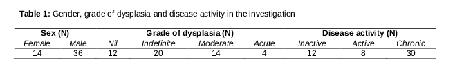
Of the 50 patients with UC studied, acute, moderate, indefinite, and no epithelial dysplasia was found in 4, 14, 20, and 12 patients, respectively (). Evaluating the UC disease activity using the protocol developed by Geboes et al [17], 30 subjects had chronic UC, 8 were active cases, and 12 subjects had inactive UC ().
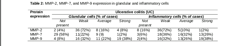
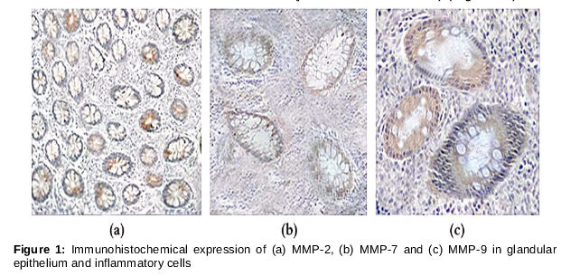
Examining glandular cells, the MMP-2 expression was weak in the glandular epithelium of 50 cases, covering 73.3 % of the patients with UC and in inflammatory cells in 72 % of the cases. MMP-2 expression in the glandular tubes was average and strong in 16 and 8 % of the cases, respectively, compared with 10 and 2 % of the inflammatory cells ().
In comparison, no MMP-7 expression was seen in the glandular epithelium of 29 patients (58 %), compared with 6 % of the inflammatory cells. There was weak expression in the glandular cells of 22 % of the cases, compared with 36 % of the inflammatory cells. MMP-9 expression was weak in both the glandular and inflammatory cells in 16 % of the cases; average expression of both was observed in 32 % of the cases ( and ).
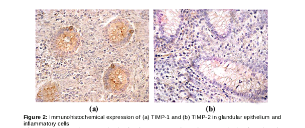
Strong TIMP-1 expression was found in the glandular epithelium in 32 cases (64 %) and in 40 % of the inflammatory infiltrate cells. There was weak TIMP-2 in glandular epithelium in 44 % of the cases, compared with 33 % of the inflammatory cells (a, b).
There was a strong correlation between the expression of MMPs in the glandular epithelium and the presence of ulcers (p = 0.051, R = 0.392). In addition, MMP-2 in the glandular epithelium tended to be correlated with a change in the tissue architecture (p = 0.076, R = 0.381) and the presence of neutrophils in the lamina propria (p = 0.077, R = 0.371) (a).
The expression of MMP-7 in the glandular epithelium was correlated with erosions (p = 0.031, R = 0.492). Decreased MMP-9 expression was observed with altered tissue architecture (p = 0.067, R = −0.392) (b, c).
The expression of MMP-2 in the lamina propria was correlated with the presence of neutrophils in an inflammatory infiltrate. In addition, the expression TIMP-1 correlated with eosinophils in the lamina propria (p = 0.024, R = 0.612) and neutrophils in the glandular epithelium (p = 0.032, R = 0.0581). There was a significant correlation with the expression of TIMP-2 in the glandular epithelium, but not with inflammatory cells (a, b).
Discussion
The expression of various metalloproteinases and their inhibitors observed in the patients with UC was related to the stage of the disease. There was weak MMP-7 expression in the glandular epithelium in 60 % of the cases with an inflammatory infiltrate. Weak MMP-2 expression was observed in the inflammatory infiltrate contrasting strong expression in the glandular epithelium, as noted by von Lampe et al [18]. MMP-2 expression was associated with erosions in the glandular epithelium. The expression of MMP-2 was also mainly associated with mesenchymal cells and neutrophils in the glandular epithelium and an inflammatory infiltrate. MMP-9 expression was observed in both the glandular epithelium and inflammatory infiltrate, as reported in patients with UC [19]. In addition, the protein activity in UC increased with the homogeneity of the inflamed mucosa.
The tissue architecture of the colon changed with the level of MMP-9 expression, implying that MMP-9 expression plays a role in the chronic inflammation of the tissue lining of the colon and remodelling of the tissue architecture. Our results revealed that MMP-9 plays a crucial role in the pathogenesis of UC, as reported elsewhere [20,21], suggesting MMP-9 as a potential target for treatment. TIMP-1 and TIMP-2 also play vital roles in regulating the activity of MMPs and maintaining equilibrium. The expression of TIMP-1 in an inflamed ulcer in UC has been reported [22].
Limitation of the study
One limitation of this study is that the tissue samples were from a Chinese population; the correlation between the expression of MMPs and TIMPs may vary in other populations of patients with UC.
Conclusion
The expression of MMPs and their inhibitors reflects UC progression. There are significant correlations between MMPs and histopatho-logical parameters. MMP-2, MMP-7 and MMP-9 are potential targets for reducing the progression of UC in patients by using inhibitors such as TIMP-1 and TIMP-2.
References
Archives


News Updates